DISCOVERIES REPORTS (ISSN 2393249X), 2022, volume 5
REVIEW ARTICLE
CITATION: Dindere ME, Bucur O. Cancer detection during surgery: FDA-approved use of pafolacianine. Discoveries Reports 2022; 5(2): e30. DOI: 10.15190/drep.2022.4
Cancer detection during surgery: FDA-approved use of pafolacianine
Mihaela Elisabeta Dindere1,2, Octavian Bucur1,3,*
1Victor Babes National Institute of Pathology, Bucharest, Romania
2Carol Davila University of Medicine and Pharmacy, Bucharest, Romania
3Viron Molecular Medicine Institute, Boston, MA, USA
* Corresponding author: Octavian Bucur, MD, PhD, Next Generation Pathology Group, Victor Babes National Institute of Pathology, Bucharest, Romania and Viron Molecular Medicine Institute, Boston, MA 02108, USA; octavian.bucur@ivb.ro; octavian.bucur@gmail.com
Abstract
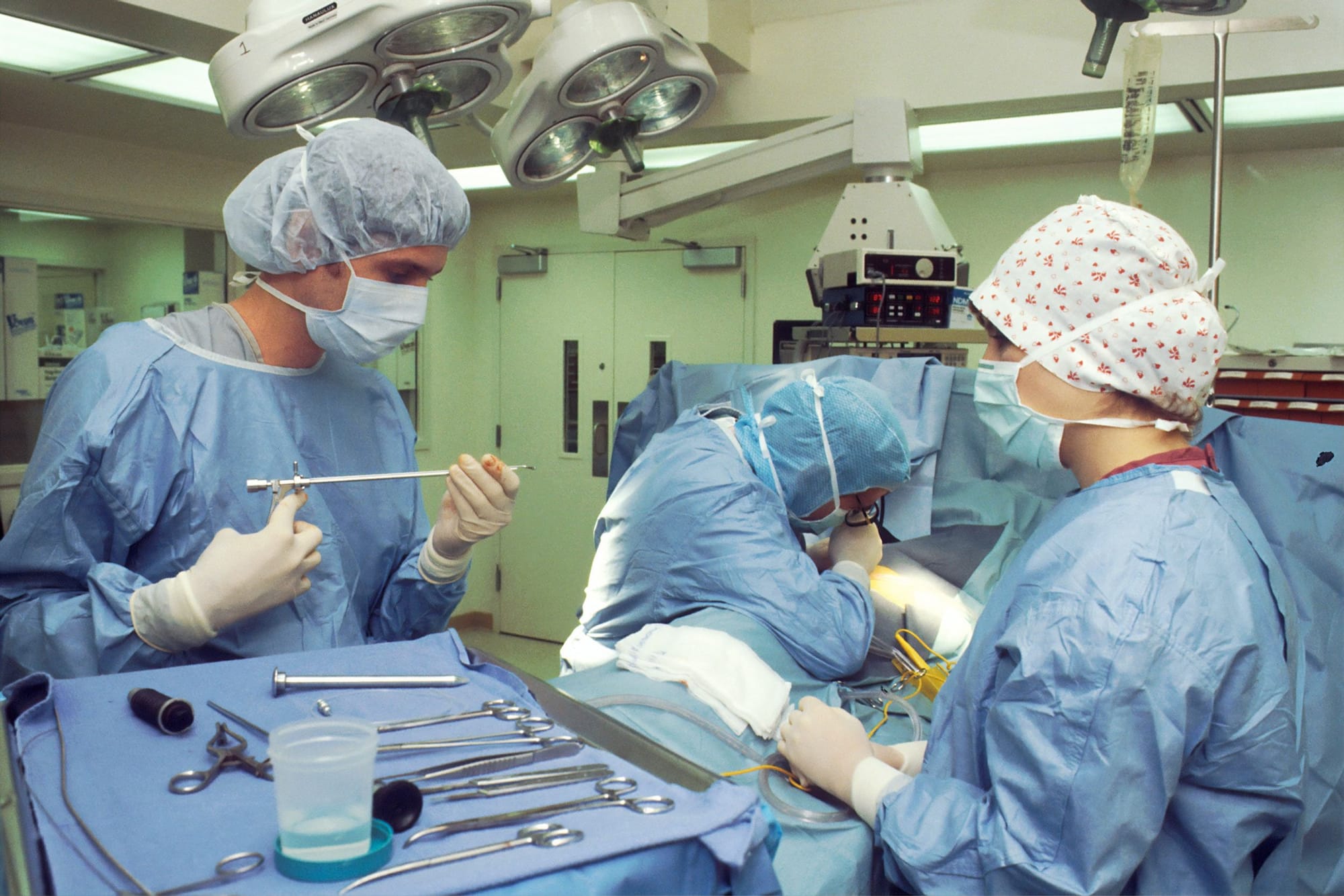
While cancer is on the rise, surgeons have limited reliable tools for intraoperative tumor detection and real-time margin assessment. The incomplete resection of the malignant lesions often leads to increased recurrence rate and ulterior interventions, as the surgeon must identify the cancer tissues mostly by inspection and palpation. Currently, the progress in finding novel tumor-specific tracers used for intraoperative molecular imaging came just in time with the advances of surgery towards minimally invasive operative approaches. Therefore, the FDA approval of pafolacianine, a FRα binding fluorescent agent used for near-infrared imaging in ovarian cancer patients, represents a step closer to achieving radical tumor excision and improved overall outcomes in oncologic patients. To date, numerous studies tested the safety and efficacy of intraoperative molecular imaging with pafolaciane in several types of cancer, but rigorous results are expected to ascertain these optimistic findings in the near future. This review highlights the beneficial use of intraoperative near-infrared imaging with pafolacianine in a wide variety of malignancies, this new developed technique succeeding in improving long-term outcomes in cancer patients by providing enriched tumor detection during surgery, and ultimately, by reducing cancer relapse rate, morbidity and mortality, costs and most importantly, the negative impact on patient survival.
Access FULL text of the manuscript here: FULL ARTICLE (PDF)
REFERENCES
1. Siegel RL, Miller KD, Fuchs HE, Jemal A. Cancer statistics, 2022. CA Cancer J Clin. 2022;72(1):7–33.
2. Surgery in cancer treatment. Canadian Cancer Society. [cited 2022 Aug 25]. Available from: https://cancer.ca/en/treatments/treatment-types/surgery
3. Hoogstins CES, Tummers QRJG, Gaarenstroom KN, De Kroon CD, Trimbos JBMZ, Bosse T, et al. A novel tumor-specific agent for intraoperative near-infrared fluorescence imaging: A translational study in healthy volunteers and patients with ovarian cancer. Clin Cancer Res. 2016;22(12):2929–38.
4. Pelosci A. A Look Behind Development of Pafolacianine for Tumor Detection During Ovarian Cancer Surgery. cancernetwork. [cited 2022 Aug 25]. Available from: https://www.cancernetwork.com/view/a-look-behind-development-of-pafolacianine-for-tumor-detection-during-ovarian-cancer-surgery
5. Cytalux (pafolacianine) for Identification of Ovarian Cancer. Clin Trials Arena. [cited 2022 Sept 25]. Available from:
https://www.clinicaltrialsarena.com/projects/cytalux-pafolacianine-for-identification-of-ovarian-cancer/
6. FDA approves pafolacianine for identifying malignant ovarian cancer lesions. U.S. Food and Drug Administration (FDA). [cited 2022 Aug 25]. Available: https://www.fda.gov/drugs/resources-information-approved-drugs/fda-approves-pafolacianine-identifying-malignant-ovarian-cancer-lesions
7. Pafolacianine. National Library of Medicine. [cited 2022 Aug 25]. Available from: https://pubchem.ncbi.nlm.nih.gov/compound/Pafolacianine
8. Pafolacianine. Drugbank. [cited 2022 Aug 25]. Available:: https://go.drugbank.com/drugs/DB15413
9. Cho SS, Lee JYK. Intraoperative Fluorescent Visualization of Pituitary Adenomas. Neurosurg Clin N Am. 2019;30(4):401–12.
10. De Jesus E, Keating JJ, Kularatne SA, Jiang J, Judy R, Predina J, et al. Comparison of Folate Receptor Targeted Optical Contrast Agents for Intraoperative Molecular Imaging. Int J Mol Imaging. 2015;2015: 469047.
11. Zhang RR, Schroeder AB, Grudzinski JJ, Rosenthal EL, Warram JM, Pinchuk AN, et al. Beyond the margins: Real-time detection of cancer using targeted fluorophores. Nat Rev Clin Oncol. 2017;14(6):347–64.
12. Birrer MJ, Betella I, Martin LP, Moore KN. Is Targeting the Folate Receptor in Ovarian Cancer Coming of Age? Oncologist. 2019;24(4):425–9.
13. Xia W, Low PS. Folate-targeted therapies for cancer. J Med Chem. 2010;53(19):6811–24.
14. O’Shannessy DJ, Yu G, Smale R, Fu YS, Singhal S, Thiel RP, et al. Folate receptor alpha expression in lung cancer: diagnostic and prognostic significance. Oncotarget. 2012 Apr;3(4):414–25.
15. Boogerd LSF, Boonstra MC, Beck AJ, Charehbili A, Hoogstins CES, Prevoo HAJM, et al. Concordance of folate receptor-α expression between biopsy, primary tumor and metastasis in breast cancer and lung cancer patients. Oncotarget. 2016 Apr;7(14):17442–54.
16. Cheung A, Bax HJ, Josephs DH, Ilieva KM, Pellizzari G, Opzoomer J, et al. Targeting folate receptor alpha for cancer treatment. Oncotarget. 2016 Aug;7(32):52553–74.
17. Newton AD, Predina JD, Frenzel-Sulyok LG, Low PS, Singhal S, Roses RE. Intraoperative Molecular Imaging Utilizing a Folate Receptor-Targeted Near-Infrared Probe Can Identify Macroscopic Gastric Adenocarcinomas. Mol Imaging Biol. 2021;23(1):11–7.
18. Predina JD, Newton AD, Connolly C, Dunbar A, Baldassari M, Deshpande C, et al. Identification of a Folate Receptor-Targeted Near-Infrared Molecular Contrast Agent to Localize Pulmonary Adenocarcinomas. Mol Ther. 2018;26(2):390–403.
19. Kalli KR, Oberg AL, Keeney GL, Christianson TJH, Low PS, Knutson KL, et al. Folate receptor alpha as a tumor target in epithelial ovarian cancer. Gynecol Oncol. 2008;108(3):619–26.
20. Markert S, Lassmann S, Gabriel B, Klar M, Werner M, Gitsch G, et al. Alpha-folate receptor expression in epithelial ovarian carcinoma and non-neoplastic ovarian tissue. Anticancer Res. 2008;28(6 A):3567–72.
21. Low PS, Kularatne SA. Folate-targeted therapeutic and imaging agents for cancer. Curr Opin Chem Biol. 2009 Jun;13(3):256–62.
22. Ross JF, Chaudhuri PK, Ratnam M. Differential regulation of folate receptor isoforms in normal and malignant tissues in vivo and in established cell lines. Physiologic and clinical implications. Cancer. 1994 May;73(9):2432–43.
23. Sulek JE, Steward JE, Bahler CD, Jacobsen MH, Sundaram A, Shum CF, et al. Folate-targeted intraoperative fluorescence, OTL38, in robotic-assisted laparoscopic partial nephrectomy. Scand J Urol. 2021;55(4):331–6.
24. Cai L, Michelakos T, Ferrone CR, Zhang L, Deshpande V, Shen Q, et al. Expression status of folate receptor alpha is a predictor of survival in pancreatic ductal adenocarcinoma. Oncotarget. 2017 Jun;8(23):37646–56.
25. Senol S, Ceyran AB, Aydin A, Zemheri E, Ozkanli S, Kösemetin D, et al. Folate receptor α expression and significance in endometrioid endometrium carcinoma and endometrial hyperplasia. Int J Clin Exp Pathol. 2015;8(5):5633–41.
26. Mahalingam SM, Kularatne SA, Myers CH, Gagare P, Norshi M, Liu X, et al. Evaluation of Novel Tumor-Targeted Near-Infrared Probe for Fluorescence-Guided Surgery of Cancer. J Med Chem. 2018 Nov;61(21):9637–46.
27. Torre LA, Trabert B, DeSantis CE, Miller KD, Samimi G, Runowicz CD, et al. Ovarian cancer statistics, 2018. CA Cancer J Clin. 2018;68(4):284–96.
28. Wentzensen N, Poole EM, Trabert B, White E, Arslan AA, Patel A V, et al. Ovarian Cancer Risk Factors by Histologic Subtype: An Analysis From the Ovarian Cancer Cohort Consortium. J Clin Oncol Off J Am Soc Clin Oncol. 2016 Aug;34(24):2888–98.
29. Kurman RJ, Shih IM. The Dualistic Model of Ovarian Carcinogenesis: Revisited, Revised, and Expanded. Am J Pathol. 2016 Apr;186(4):733–47.
30. Pearce CL, Rossing MA, Lee AW, Ness RB, Webb PM, Chenevix-Trench G, et al. Combined and interactive effects of environmental and GWAS-identified risk factors in ovarian cancer. Cancer Epidemiol biomarkers Prev a Publ Am Assoc Cancer Res cosponsored by Am Soc Prev Oncol. 2013 May;22(5):880–90.
31. Prat J. New insights into ovarian cancer pathology. Vol. 23 Suppl 10, Annals of oncology : official journal of the European Society for Medical Oncology. England; 2012. p. x111-7.
32. Van Dam GM, Themelis G, Crane LMA, Harlaar NJ, Pleijhuis RG, Kelder W, et al. Intraoperative tumor-specific fluorescence imaging in ovarian cancer by folate receptor-α targeting: First in-human results. Nat Med. 2011;17(10):1315–9.
33. Chang SJ, Bristow RE, Ryu HS. Impact of complete cytoreduction leaving no gross residual disease associated with radical cytoreductive surgical procedures on survival in advanced ovarian cancer. Ann Surg Oncol. 2012;19(13):4059–67.
34. Vergote I, Tropé CG, Amant F, Kristensen GB, Ehlen T, Johnson N, et al. Neoadjuvant Chemotherapy or Primary Surgery in Stage IIIC or IV Ovarian Cancer. N Engl J Med. 2010;363(10):943–53.
35. Bristow RE, Tomacruz RS, Armstrong DK, Trimble EL, Montz FJ. Survival effect of maximal cytoreductive surgery for advanced ovarian carcinoma during the platinum era: A meta-analysis. J Clin Oncol. 2002;20(5):1248–59.
36. Hoskins WJ, McGuire WP, Brady MF, Homesley HD, Creasman WT, Berman M, et al. The effect of diameter of largest residual disease on survival after primary. AmJObstetGynecol. 1994;170(4):974–9.
37. Bristow RE, Berek JS. Surgery for ovarian cancer: how to improve survival. Lancet. 2006;367(9522):1558–60.
38. Shen J, Hilgenbrink AR, Xia W, Feng Y, Dimitrov DS, Lockwood MB, et al. Folate receptor-β constitutes a marker for human proinflammatory monocytes. J Leukoc Biol. 2014;96(4):563–70.
39. O’Shannessy DJ, Somers EB, Wang LC, Wang H, Hsu R. Expression of folate receptors alpha and beta in normal and cancerous gynecologic tissues: Correlation of expression of the beta isoform with macrophage markers. J Ovarian Res. 2015;8(1):1–9.
40. Puig-Kröger A, Sierra-Filardi E, Domínguez-Soto A, Samaniego R, Corcuera MT, Gómez-Aguado F, et al. Folate receptor β is expressed by tumor-associated macrophages and constitutes a marker for M2 anti-inflammatory/regulatory Macrophages. Cancer Res. 2009;69(24):9395–403.
41. Kurahara H, Takao S, Kuwahata T, Nagai T, Ding Q, Maeda K, et al. Clinical Significance of Folate Receptor β–expressing Tumor-associated Macrophages in Pancreatic Cancer. Ann Surg Oncol. 2012;19(7):2264–71.
42. Smith HA, Kang Y. The metastasis-promoting roles of tumor-associated immune cells. J Mol Med. 2013;91(4):411–29.
43. Joyce JA, Pollard JW. Microenvironmental regulation of metastasis. Nat Rev Cancer. 2009;9(4):239–52.
44. Lewis CE, Pollard JW. Distinct role of macrophages in different tumor microenvironments. Cancer Res. 2006;66(2):605–12.
45. Keereweer S, Van Driel PBAA, Snoeks TJA, Kerrebijn JDF, De Jong RJB, Vahrmeijer AL, et al. Optical image-guided cancer surgery: Challenges and limitations. Clin Cancer Res. 2013;19(14):3745–54.
46. Weissleder R, Ntziachristos V. Shedding light onto live molecular targets. Nat Med. 2003;9(1):123–8.
47. Monici M. Cell and tissue autofluorescence research and diagnostic applications. Biotechnol Annu Rev. 2005;11:227–56.
48. Monici M, Basile V, Romano G, Evangelisti L, Lucarini L, Attanasio M, et al. Fibroblast autofluorescence in connective tissue disorders: a future tool for clinical and differential diagnosis? J Biomed Opt. 2008;13(5):54025.
49. Rosenthal EL, Warram JM, de Boer E, Chung TK, Korb ML, Brandwein-Gensler M, et al. Safety and Tumor Specificity of Cetuximab-IRDye800 for Surgical Navigation in Head and Neck Cancer. Clin cancer Res an Off J Am Assoc Cancer Res. 2015 Aug;21(16):3658–66.
50. Randall LM, Wenham RM, Low PS, Dowdy SC, Tanyi JL. A phase II, multicenter, open-label trial of OTL38 injection for the intra-operative imaging of folate receptor-alpha positive ovarian cancer. Gynecol Oncol. 2019;155(1):63–8.
51. Lung cancer statistics. World Cancer Research Fund International. [cited 2022 Sept 25]. Available from: https://www.wcrf.org/cancer-trends/lung-cancer-statistics/
52. Facts about lung cancer. Lung Cancer Research Foundation. Available from: https://www.lungcancerresearchfoundation.org/lung-cancer-facts/
53. Non-Small Cell Lung Cancer. Yale Medicine. [cited 2022 Aug 25]. Available from: https://www.yalemedicine.org/conditions/non-small-cell-lung-cancer
54. Cerfolio RJ, Bryant AS. Is palpation of the nonresected pulmonary lobe(s) required for patients with non-small cell lung cancer? A prospective study. J Thorac Cardiovasc Surg. 2008;135(2):261–8.
55. Predina JD, Newton A, Corbett C, Xia L, Sulyok LF, Shin M, et al. Localization of Pulmonary Ground-Glass Opacities with Folate Receptor–Targeted Intraoperative Molecular Imaging. J Thorac Oncol. 2018;13(7):1028–36.
56. Keating J, Singhal S. Novel Methods of Intraoperative Localization and Margin Assessment of Pulmonary Nodules. Semin Thorac Cardiovasc Surg. 2016;28(1):127–36.
57. Park CH, Han K, Hur J, Lee SM, Lee JW, Hwang SH, et al. Comparative Effectiveness and Safety of Preoperative Lung Localization for Pulmonary Nodules: A Systematic Review and Meta-analysis. Chest. 2017 Feb;151(2):316–28.
58. Choi NK, Solomon DH, Tsacogianis TN, Landon JE, Song HJ, Kim SC. Comparative Safety and Effectiveness of Denosumab Versus Zoledronic Acid in Patients With Osteoporosis: A Cohort Study. J bone Miner Res Off J Am Soc Bone Miner Res. 2017 Mar;32(3):611–7.
59. Zaman M, Bilal H, Woo CY, Tang A. In patients undergoing video-assisted thoracoscopic surgery excision, what is the best way to locate a subcentimetre solitary pulmonary nodule in order to achieve successful excision? Interact Cardiovasc Thorac Surg. 2012 Aug;15(2):266–72.
60. Gao JW, Rizzo S, Ma LH, Qiu XY, Warth A, Seki N, et al. Pulmonary ground-glass opacity: Computed tomography features, histopathology and molecular pathology. Transl Lung Cancer Res. 2017;6(1):68–75.
61. Tipirneni KE, Warram JM, Moore LS, Prince AC, De Boer E, Jani AH, et al. Oncologic Procedures Amenable to Fluorescence-guided Surgery. Ann Surg. 2017;266(1):36–47.
62. Tipirneni KE, Rosenthal EL, Moore LS, Haskins AD, Udayakumar N, Jani AH, et al. Fluorescence Imaging for Cancer Screening and Surveillance. Mol Imaging Biol. 2017;19(5):645–55.
63. Newton AD, Predina JD, Nie S, Low PS, Singhal S. Intraoperative fluorescence imaging in thoracic surgery. J Surg Oncol. 2018 Aug;118(2):344–55.
64. Colombo N, Creutzberg C, Amant F, Bosse T, González-Martín A, Ledermann J, et al. ESMO-ESGO-ESTRO Consensus Conference on Endometrial Cancer: diagnosis, treatment and follow-up. Ann Oncol Off J Eur Soc Med Oncol. 2016 Jan;27(1):16–41.
65. Wu M, Gunning W, Ratnam M. Expression of folate receptor type alpha in relation to cell type, malignancy, and differentiation in ovary, uterus, and cervix. Cancer Epidemiol biomarkers Prev a Publ Am Assoc Cancer Res cosponsored by Am Soc Prev Oncol. 1999 Sep;8(9):775–82.
66. Shen J, Putt KS, Visscher DW, Murphy L, Cohen C, Singhal S, et al. Assessment of folate receptor-β expression in human neoplastic tissues. Oncotarget. 2015;6(16):14700–9.
67. Boogerd LSF, Hoogstins CES, Gaarenstroom KN, de Kroon CD, Beltman JJ, Bosse T, et al. Folate receptor-α targeted near-infrared fluorescence imaging in high-risk endometrial cancer patients: a tissue microarray and clinical feasibility study. Oncotarget. 2018 Jan;9(1):791–801.
68. Parker N, Turk MJ, Westrick E, Lewis JD, Low PS, Leamon CP. Folate receptor expression in carcinomas and normal tissues determined by a quantitative radioligand binding assay. Anal Biochem. 2005;338(2):284–93.
69. Shum CF, Bahler CD, Low PS, Ratliff TL, Kheyfets S V., Natarajan JP, et al. Novel Use of Folate-Targeted Intraoperative Fluorescence, OTL38, in Robot-Assisted Laparoscopic Partial Nephrectomy: Report of the First Three Cases. J Endourol Case Reports. 2016;2(1):189–97.
70. Hekman MCH, Rijpkema M, Langenhuijsen JF, Boerman OC, Oosterwijk E, Mulders PFA. Intraoperative Imaging Techniques to Support Complete Tumor Resection in Partial Nephrectomy. Eur Urol Focus. 2018 Dec;4(6):960–8.
71. Guzzo TJ, Jiang J, Keating J, DeJesus E, Judy R, Nie S, et al. Intraoperative Molecular Diagnostic Imaging Can Identify Renal Cell Carcinoma. J Urol. 2016 Mar;195(3):748–55.
72. Wallis CJD, Garbens A, Chopra S, Gill IS, Satkunasivam R. Robotic Partial Nephrectomy: Expanding Utilization, Advancing Innovation. J Endourol. 2017 Apr;31(4):348–54.
73. Ghani KR, Sukumar S, Sammon JD, Rogers CG, Trinh QD, Menon M. Practice patterns and outcomes of open and minimally invasive partial nephrectomy since the introduction of robotic partial nephrectomy: results from the nationwide inpatient sample. J Urol. 2014 Apr;191(4):907–12.
74. Rogers CG, Laungani R, Bhandari A, Krane LS, Eun D, Patel MN, et al. Maximizing console surgeon independence during robot-assisted renal surgery by using the Fourth Arm and TilePro. J Endourol. 2009 Jan;23(1):115–21.
75. Krane LS, Manny TB, Hemal AK. Is near infrared fluorescence imaging using indocyanine green dye useful in robotic partial nephrectomy: a prospective comparative study of 94 patients. Urology. 2012 Jul;80(1):110–6.
76. Amin MB, Greene FL, Edge SB, Compton CC, Gershenwald JE, Brookland RK, et al. The Eighth Edition AJCC Cancer Staging Manual: Continuing to build a bridge from a population-based to a more “personalized” approach to cancer staging. Vol. 67, CA: a cancer journal for clinicians. United States; 2017. p. 93–9.
77. Ajani JA, D’Amico TA, Bentrem DJ, Chao J, Cooke D, Corvera C, et al. Gastric Cancer, Version 2.2022, NCCN Clinical Practice Guidelines in Oncology. J Natl Compr Canc Netw. 2022 Feb;20(2):167–92.
78. Bentrem D, Gerdes H, Tang L, Brennan M, Coit D. Clinical correlation of endoscopic ultrasonography with pathologic stage and outcome in patients undergoing curative resection for gastric cancer. Ann Surg Oncol. 2007 Jun;14(6):1853–9.
79. Spolverato G, Ejaz A, Kim Y, Squires MH, Poultsides GA, Fields RC, et al. Use of endoscopic ultrasound in the preoperative staging of gastric cancer: a multi-institutional study of the US gastric cancer collaborative. J Am Coll Surg. 2015 Jan;220(1):48–56.
80. Xi WD, Zhao C, Ren GS. Endoscopic ultrasonography in preoperative staging of gastric cancer: determination of tumor invasion depth, nodal involvement and surgical resectability. World J Gastroenterol. 2003 Feb;9(2):254–7.
81. Bucur O. Emerging technologies for diagnostic pathology. Discoveries. 2015 Jun;3(2):e46.
82. Bucur O, Fu F, Calderon M, Mylvaganam GH, Ly NL, Day J, et al. Nanoscale imaging of clinical specimens using conventional and rapid-expansion pathology. Nat Protoc. 2020 May;15(5):1649–72.
83. Mediu R, Rama A, Mediu N. Screening for prostate cancer: a study on the free and total prostate specific antigen. Discoveries. 2021;9(4):e139.
84. Klimas A, Bucur O, Njeri B, Zhao Y. Nanoscopic Imaging of Human Tissue Sections via Physical and Isotropic Expansion. J Vis Exp. 2019 Sep;(151).
85. Nishith N, Rao RN, Rai P. Cytologic Categorization with Risk Stratification of Endoscopic Ultrasound-Guided Fine Needle Aspiration from Pancreatic Lesions Based on Guidelines of the Papanicolaou Society of Cytopathology: 12-Year Tertiary Care Experience. Discoveries. 2021;9(3):e134.
86. Zhao Y, Bucur O, Irshad H, Chen F, Weins A, Stancu AL, et al. Nanoscale imaging of clinical specimens using pathology-optimized expansion microscopy. Nat Biotechnol. 2017 Aug;35(8):757–64.
87. Lee CM, Tian X, Tsao C, Chen P, Huang TN, Hsueh YP, et al. Macro Photography with Lightsheet Illumination Enables Whole Expanded Brain Imaging with Single-cell Resolution. Discoveries. 2021;9(3):e133.
88. Predina JD, Newton AD, Keating J, Barbosa EM, Okusanya O, Xia L, et al. Intraoperative Molecular Imaging Combined with Positron Emission Tomography Improves Surgical Management of Peripheral Malignant Pulmonary Nodules. Ann Surg. 2017;266(3):479–88.
89. Galt JR, Halkar RK, Evans CO, Osman NA, LaBorde D, Fox TH, et al. In vivo assay of folate receptors in nonfunctional pituitary adenomas with99mTc-Folate SPECT/CT. J Nucl Med. 2010;51(11):1716–23.
90. Evans CO, Yao C, LaBorde D, Oyesiku NM. Chapter 8 Folate Receptor Expression in Pituitary Adenomas. Cellular and Molecular Analysis. Vitam Horm. 2008;79(08):235–66.
91. Evans CO, Reddy P, Brat DJ, O’Neill EB, Craige B, Stevens VL, et al. Differential expression of folate receptor in pituitary adenomas. Cancer Res. 2003 Jul;63(14):4218–24.
92. Evans CO, Young AN, Brown MR, Brat DJ, Parks JS, Neish AS, et al. Novel patterns of gene expression in pituitary adenomas identified by complementary deoxyribonucleic acid microarrays and quantitative reverse transcription-polymerase chain reaction. J Clin Endocrinol Metab. 2001 Jul;86(7):3097–107.
93. Lee JYK, Cho SS, Zeh R, Pierce JT, Martinez-Lage M, Adappa ND, et al. Folate receptor overexpression can be visualized in real time during pituitary adenoma endoscopic transsphenoidal surgery with near-infrared imaging. J Neurosurg. 2018;129(2):390–403.
94. Cho SS, Jeon J, Buch L, Nag S, Nasrallah M, Low PS, et al. Intraoperative near-infrared imaging with receptor-specific versus passive delivery of fluorescent agents in pituitary adenomas. J Neurosurg. 2019;131(6):1974–84.
95. Cho SS, Zeh R, Pierce JT, Jeon J, Nasrallah M, Adappa ND, et al. Folate Receptor Near-Infrared Optical Imaging Provides Sensitive and Specific Intraoperative Visualization of Nonfunctional Pituitary Adenomas. Oper Neurosurg. 2019;16(1):59–70.