DISCOVERIES REPORTS (ISSN 2393249X), 2023, volume 6
ORIGINAL ARTICLE
CITATION: Fortner A. RNA seqFISH: A High-Resolution Method for Spatial Transcriptomics. Discoveries Reports 2023; 6: e41. DOI: 10.15190/drep.2023.5
RNA seqFISH: A High-Resolution Method for Spatial Transcriptomics
Andra Fortner 1, *, Octavian Bucur 2,3 *
1 Medical School, Ruprecht-Karls-Universität Heidelberg, 69120 Heidelberg, Germany
2 Genomics Research and Development Institute, Bucharest, Romania
3 Viron Molecular Medicine Institute, Boston, MA 02108, USA
* Corresponding authors:
Andra Fortner, Medical School, Ruprecht-Karls-Universität Heidelberg, 69120 Heidelberg, Germany; E-mail: fortnerandra@gmail.com; Octavian Bucur, Genomics Research and Development Institute, Bucharest, Romania; Email: octavian.bucur@genomica.gov.ro
Abstract
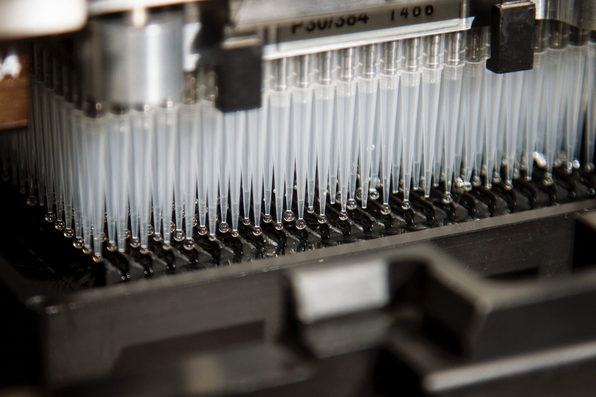
By revealing the molecular processes and interactions within cells, omics technologies have transformed our comprehension of cellular functions and metabolism. RNA seqFISH is a spatial transcriptomics method that enables high-resolution imaging of thousands of genes in single cells within tissues, revealing the spatial organization and dynamics of the transcript One in situ. In this article, we review the method itself and the applications of this technology in various biological contexts, such as development, differentiation, and disease. We discuss the benefits of the RNA seqFISH technique to provide us new insights into the molecular mechanisms underlying cellular function and diversity, as well as the limitations of this technique. We conclude that RNA seqFISH is a powerful and promising tool for studying spatial transcriptomics, and that it will inspire more researchers to adopt and advance this technology.
Access FULL text of the manuscript here: FULL ARTICLE (PDF)
REFERENCES
1. Raj A, van den Bogaard P, Rifkin SA, van Oudenaarden, A, Tyagi S. Imaging individual mRNA molecules using multiple singly labeled probes. Nat Methods. 2008; 5: 877.
2. Chen J, McSwiggen D, Ünal E. Single Molecule Fluorescence in Situ Hybridization (smFISH) Analysis in Budding Yeast Vegetative Growth and Meiosis. J Vis Exp. 2018, 57774.
3. Lubeck E, Coskun AF, Zhiyentayev T, Ahmad M, Cai L. Single cell in situ RNA profiling by sequential hybridization. Nat Methods. 2014; 11: 360.
4. Shah S, Lubeck E, Zhou W, Cai L. In Situ Transcription Profiling of Single Cells Reveals Spatial Organization of Cells in the Mouse Hippocampus. Neuron. 2016; 92: 342–357.
5. Lubeck E, Coskun AF, Zhiyentayev T, Ahmad M Cai L. Single-cell in situ RNA profiling by sequential hybridization. Nature Methods. 2014; 11: 360–361.
6. Wu Z, Liu GQ, Yang XL, Jiang JH. Electrostatic nucleic acid nanoassembly enables hybridization chain reaction in living cells for ultrasensitive mRNA Imaging. J Am Chem Soc. 2015; 137: 6829–6836.
7. Lohoff T et al. Integration of spatial and single-cell transcriptomic data elucidates mouse organogenesis. Nat Biotechnol. 2022; 40: 74–85.
8. Zhou W et al. Single-Cell Analysis Reveals Regulatory Gene Expression Dynamics Leading to Lineage Commitment in Early T Cell Development. Cell Syst. 2019; 9: 321-337.e9.
9. Kim DW et al. Multimodal Analysis of Cell Types in a Hypothalamic Node Controlling Social Behavior. Cell. 2019; 179, 713.
10. Eng CHL et al. Transcriptome-scale super-resolved imaging in tissues by RNA seqFISH+. Nature 2019 568: 235–239.
11. Shah S et al. Dynamics and spatial genomics of the nascent transcriptome by intron seqFISH. Cell. 2018; 174: 363.
12. Dar D, Dar N, Cai L, Newman DK. Spatial transcriptomics of planktonic and sessile bacterial populations at single-cell resolution. Science. 2021; 373.
13. Vu T et al. Spatial transcriptomics using combinatorial fluorescence spectral and lifetime encoding, imaging and analysis. Nat Commun. 2022; 13.
14. Rao A, Barkley D, França GS, Yanai I. Exploring tissue architecture using spatial transcriptomics. Nature. 2021; 596: 211